Sex differences in neurodegenerative diseases associated with oxidative stress, such
as Alzheimer’s disease and Parkinson’s disease
We use neuronal and astrocyte cell lines derived from males and females to examine
the impact of the sex chromosome complement on oxidative stress damage. Gonadal hormones,
such as androgens and estrogens, are used to determine the role of sex hormones on
oxidative stress damage.
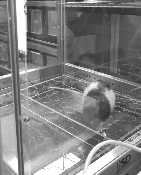
We use two different rat models to induce oxidative stress damage associated with
neurodegenerative diseases: hypoxia to induce global oxidative stress and beta-amyloid
intrahippocampal injections to induce oxidative stress damage to the brain. Using
animal models allows us to perform functional studies, such as behavior (cognition,
fine and gross motor, social interactions, ultrasonic vocalizations, olfaction behaviors).
We also use human samples via a collaboration with the Institute for Translational
Research that is a biorepository containing thousands of samples from participants
with different cognitive status and a range of ethnicities. Using these samples, we
are able to examine the impact of sex on cognition.
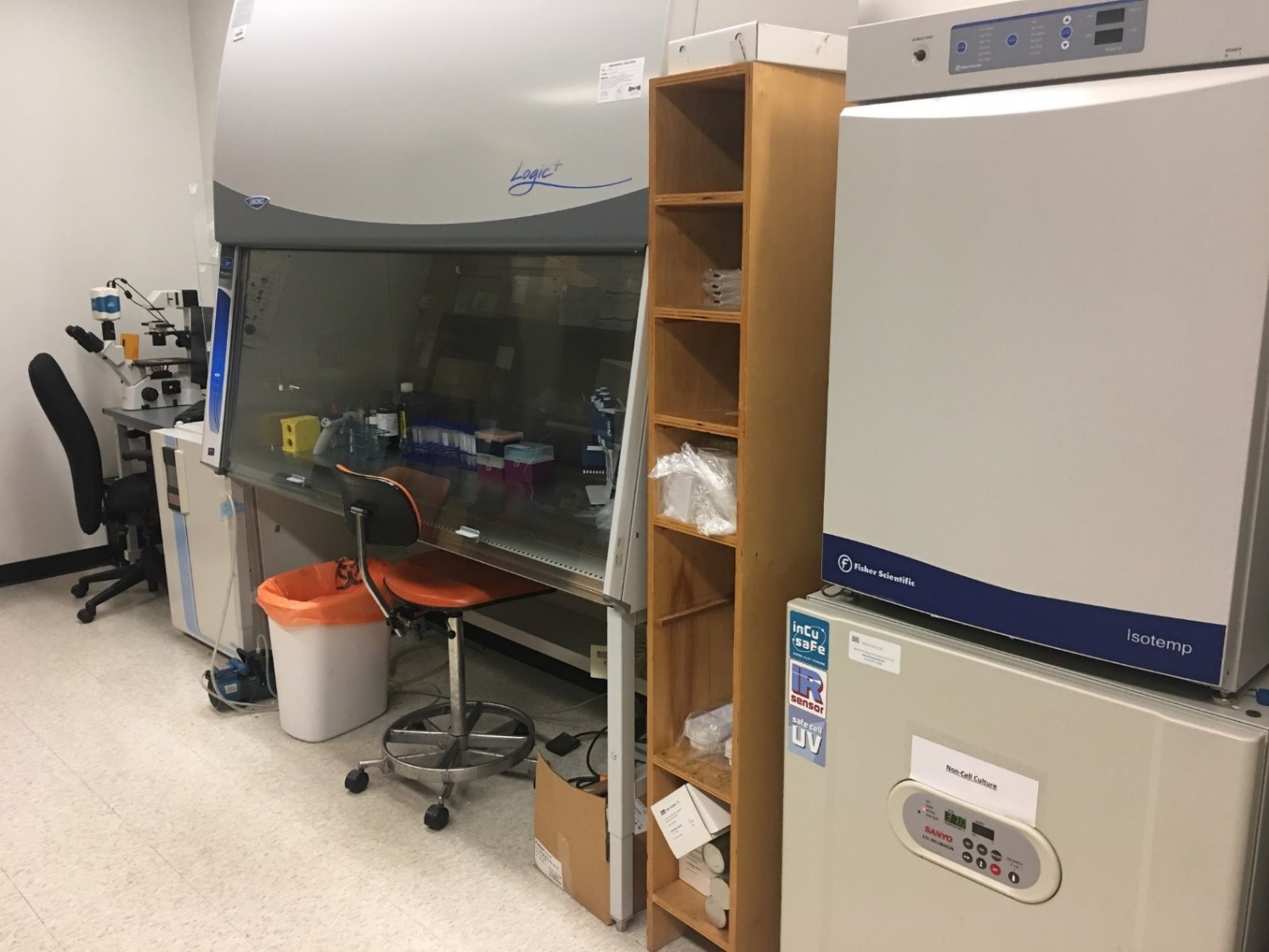
Integrative techniques used in the lab
Cell culture to examine signaling cascades
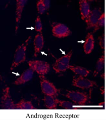
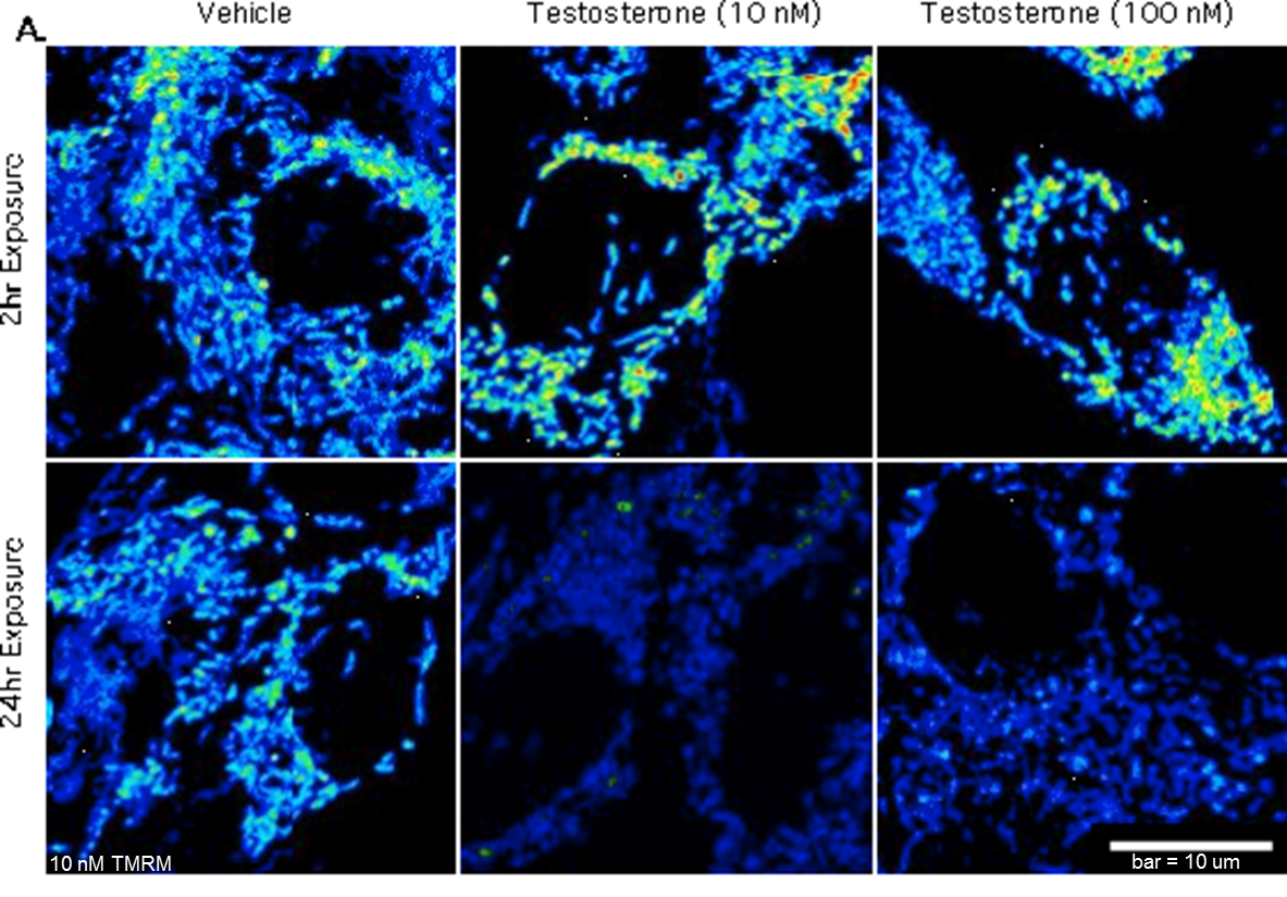
Microscopy to examine proteins and cellular integrity in cells and tissues. Real-time
microscopy to examine signaling function in cells (e.g. endocytosis, cellular hormone
release, mitochondria membrane potentials)
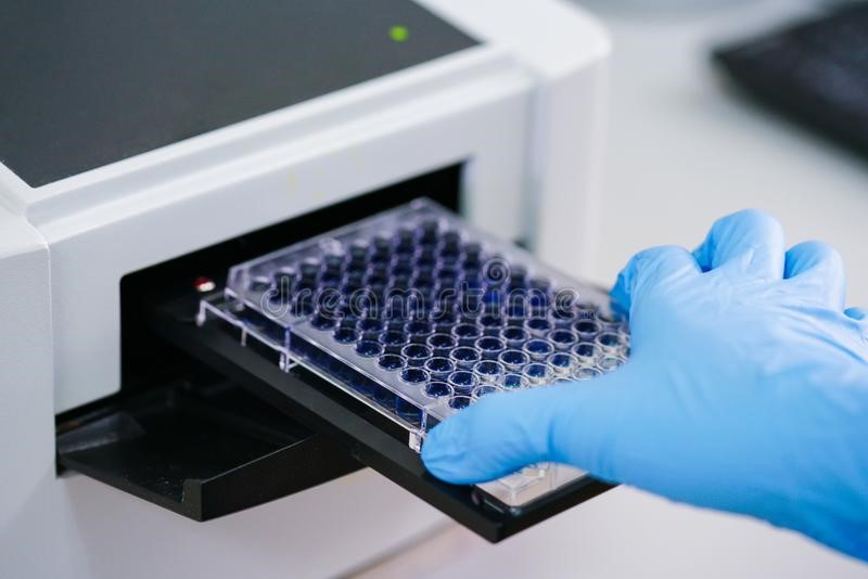
ELISAs are used to measure oxidative stress, sex hormones, gonadotropins
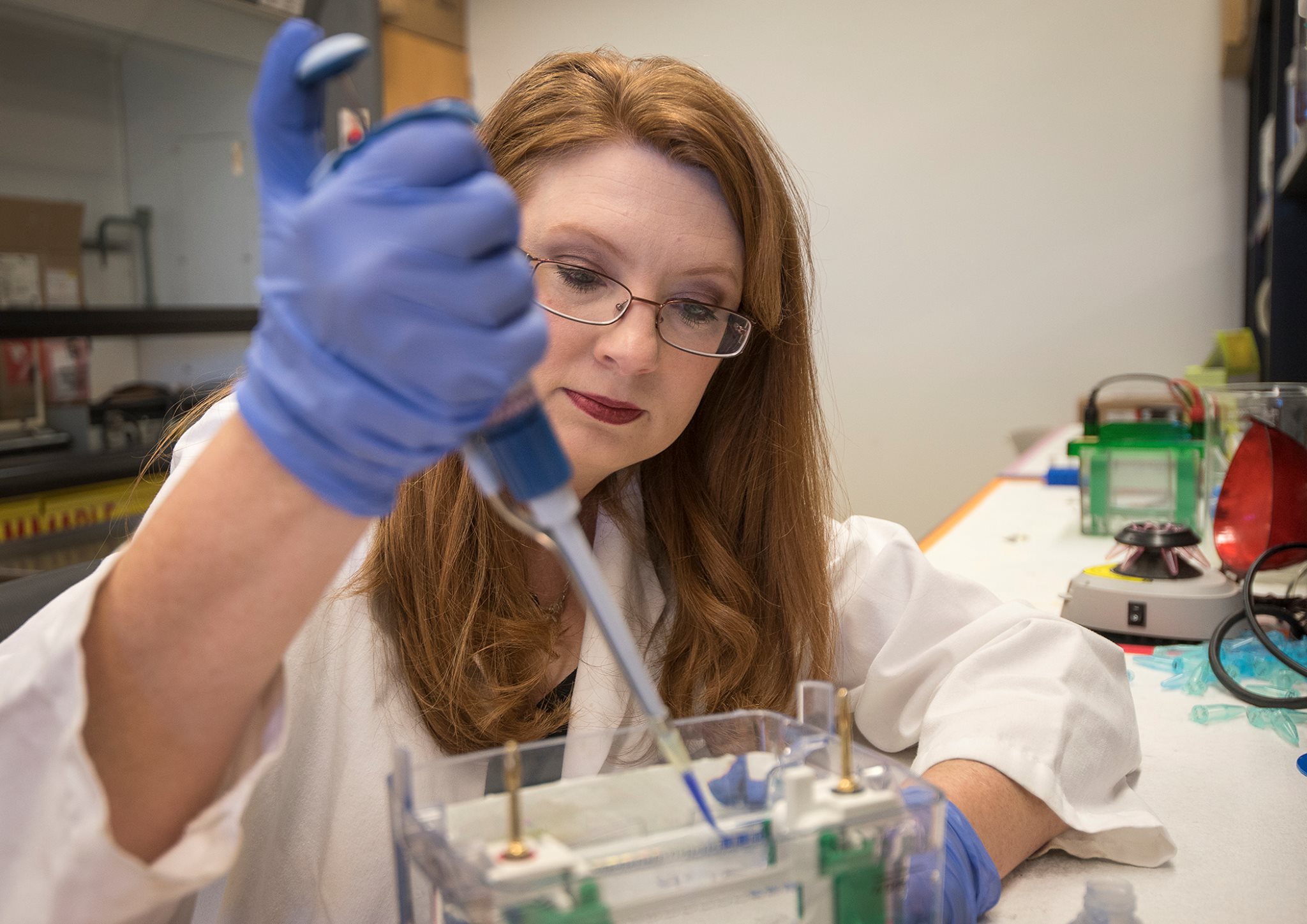
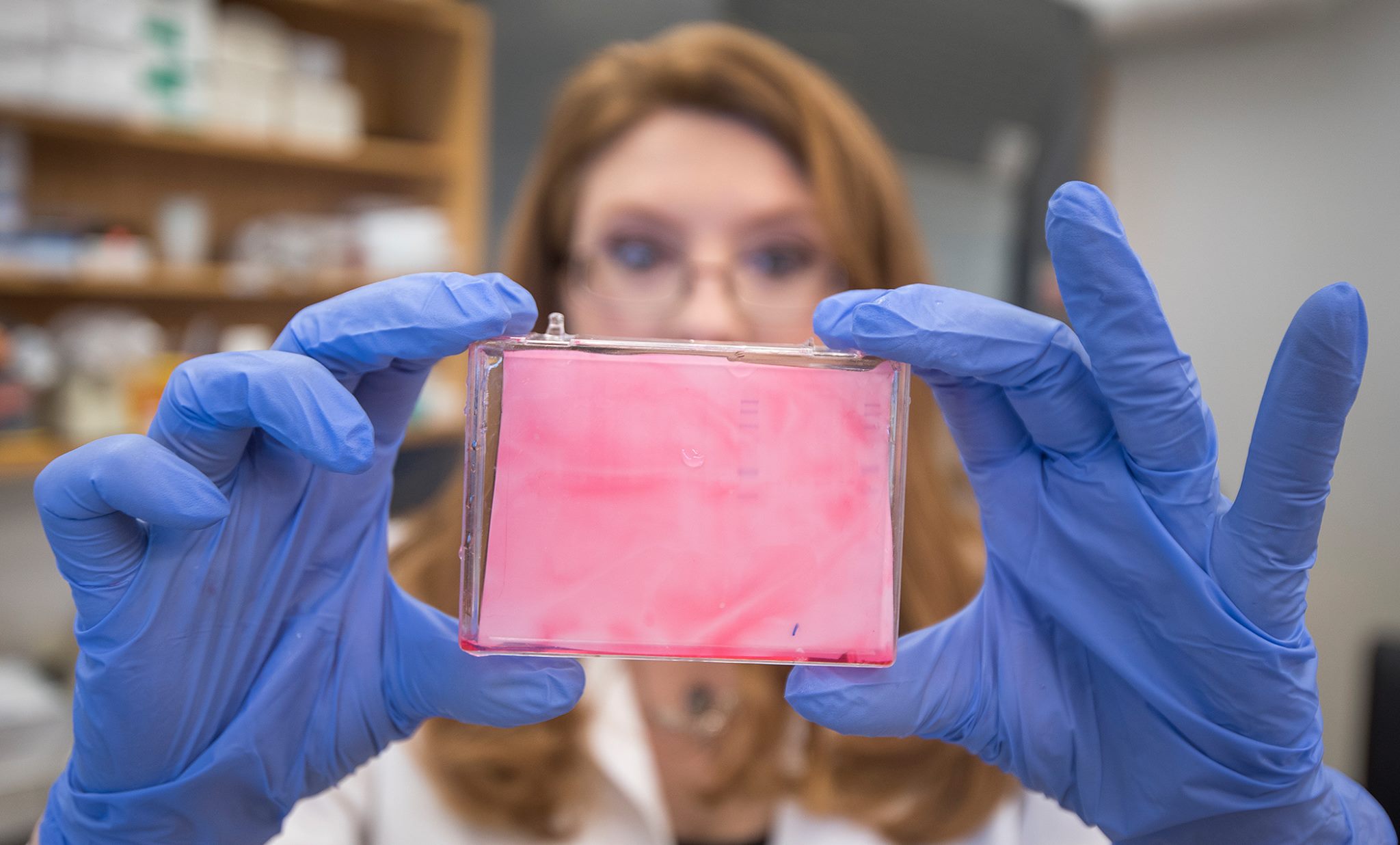
Protein analysis: electrophoresis and western blotting on whole cells, fractionated
cells (membrane, cytosol, and nuclear compartments), and co-immunoprecipitated proteins
mRNA analysis: quantitative PCR
Behavior analysis
Stereotaxic surgery to deliver agents to specific areas of the brain